Peripheral Vascular Disease Assessment
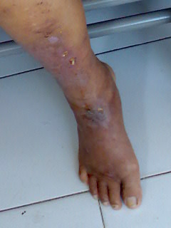
Peripheral vascular disease assessment. Comment on any items of clinical note around the bed. By a reduction in blood flow and hence 02 through the peripheral vessels when the need of the tissues for 02 exceeds the supply areas of ischemia and necrosis will develop 3. Look for signs of obvious vascular compromise.
Diabetes mellitus is a risk factor for this disease. In diabetes pain perception may be blunted by the presence of peripheral neuropathy. The peripheral vascular perfusion scan uniquely and under varied physiologic conditions permits assessment of the effect of arterial disease on the distribution of perfusion within the extremity.
Occlusive disease of the arteries of the extremity. Peripheral vascular assessment includes portions of a skin assessment as well as pulses and other indicators of perfusion. Knowing the warning signs of PAD and seeking treatment early can drastically reduce your risk of.
This article describes assessment of patients. The initial assessment is the most important part of the management of any patients management. The peripheral vascular PVS examination is performed to elicit signs of peripheral vascular pathology such as examining the blood vessels in the extremities.
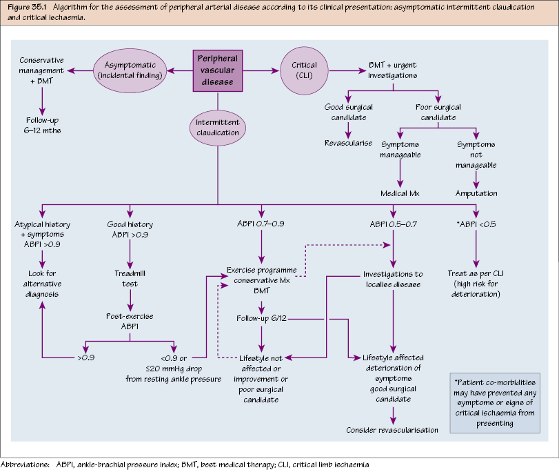
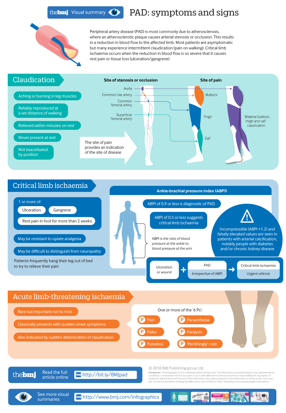
Assess the patient from the end of the bed. A differential assessment is essential to identify the type of PVD and to plan management strategies to prevent further vascular dysfunction. Peripheral vascular disease PVD is a common reason for referral to vascular clinics conditions of which include intermittent claudication and in emergency situations ischaemia of the limbs.
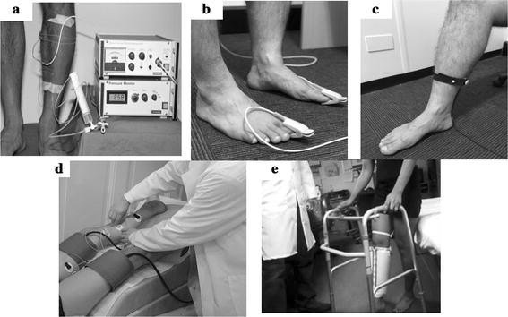
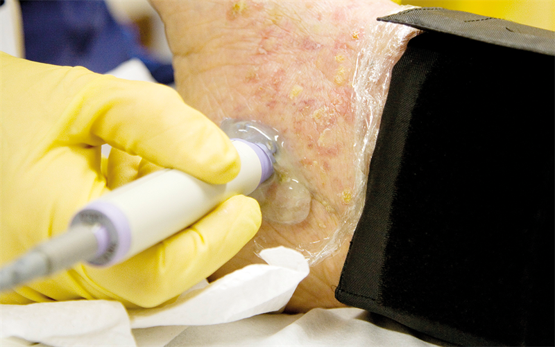
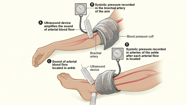
Vascular ultrasound provides a valuable tool in the assessment of various peripheral vascular beds. This is an examination using sound waves thich go through tissue and strike the RBCs and bounce back. Peripheral vascular disease assessment in the lower limb.
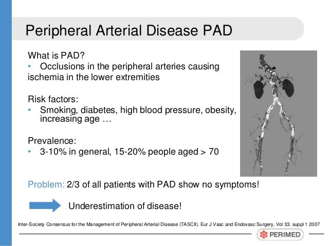
Peripheral vascular disease PVD is a chronic limb ischaemia caused by atherosclerosis of the peripheral arteries. Despite the advances in computed tomography CT and magnetic resonance MR angiographic techniques vascular ultrasound still represents the initial test of choice based on its noninvasive nature accuracy validated values and wide availability.
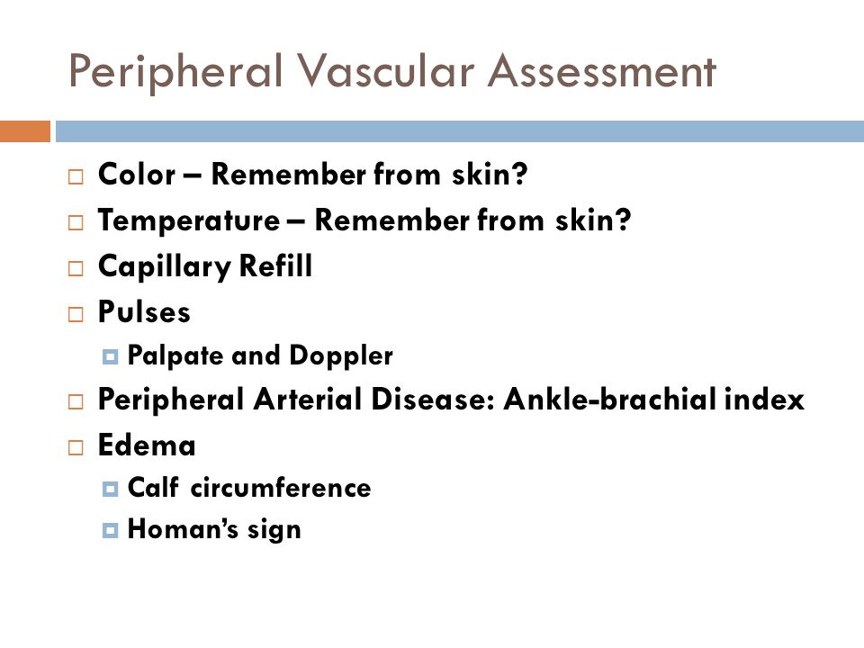
Peripheral vascular assessment includes portions of a skin assessment as well as pulses and other indicators of perfusion.
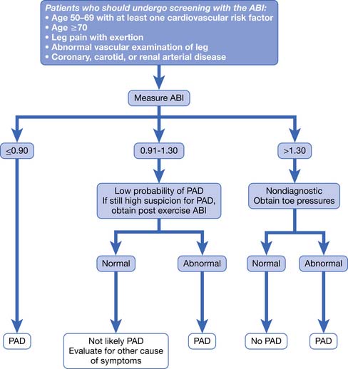
Knowledge of the range of normal and collateral anatomy is. Contents To succinctly examine the peripheral vascular system and present your findings to a consultant examiner in 6 minutes Discuss disease processes behind your clinical findings Pass the exam. Clinical Assessment for PAD History and Physical Examination Ankle-Brachial Index ABI Physiological Testing Vascular Lab Anatomic Imaging Assessment Assessing PAD Patients for Disease in Other Vascular Beds. What is PVD. The most common symptom of PVD is muscle pain in the lower limbs on exercise. The peripheral vascular PVS examination is performed to elicit signs of peripheral vascular pathology such as examining the blood vessels in the extremities. Peripheral Vascular Examination By Doctor Aram Baram MDMRCSEd 2. It evaluates blood flow and can locate a thrombus. Color of skin and nail beds.
Peripheral vascular disease PVD is a chronic limb ischaemia caused by atherosclerosis of the peripheral arteries. Clinical Assessment for PAD History and Physical Examination Ankle-Brachial Index ABI Physiological Testing Vascular Lab Anatomic Imaging Assessment Assessing PAD Patients for Disease in Other Vascular Beds. A review of current and emerging non-invasive diagnostic methods Biomed Eng Online. Peripheral artery disease or PAD affects nearly 18 million Americans and goes undiagnosed in many more. Peripheral vascular disease is categorized as either venous arterial or mixed. In diabetes pain perception may be blunted by the presence of peripheral neuropathy. 2016 AHAACC PAD Guideline.


















Post a Comment for "Peripheral Vascular Disease Assessment"